Amazing Science
1.1M views | +5 today
Follow
Amazing Science
Amazing science facts - 3D_printing • aging • AI • anthropology • art • astronomy • bigdata • bioinformatics • biology • biotech • chemistry • computers • cosmology • education • environment • evolution • future • genetics • genomics • geosciences • green_energy • language • map • material_science • math • med • medicine • microscopy • nanotech • neuroscience • paleontology • photography • photonics • physics • postings • robotics • science • technology • video
Your new post is loading...
Scooped by Dr. Stefan Gruenwald
Scoop.it!

New light technique could result in less intrusive, more effective disease diagnosis in medicine

New light technique could result in less intrusive, more effective disease diagnosis in medicine | Amazing Science | Scoop.it

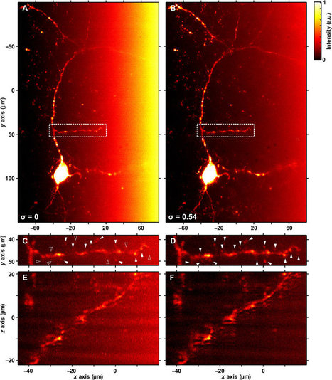
A new method of using light to scan the human body, developed by researchers at the University of St Andrews, could result in less intrusive and more effective diagnosis for patients. The work is the result of a collaboration between researchers from the Schools of Physics and Astronomy, Biology, Medicine and the Scottish Oceans Institute at the University.


The new technique allows the light to be shaped so it can reach greater depths within biological tissue enabling high quality three-dimensional (3-D) images to be acquired. It can also allow detailed 3-D images of biological specimens to be made without dissection or having to rotate specimens and take multiple images which are then fused together.


Published in the journal Science Advances, the research shows that the new method adds value to two existing imaging techniques—Bessel beam based light-sheet microscopy and Airy beam based light-sheet microscopy. Dr. Jonathan Nylk of the School of Physics and Astronomy said: "We've recently discovered particular beam shapes that retain their shape when traveling through biological tissue. These beams, called Airy beams and Bessel beams resist the effects of scattering but they still become dimmer as they travel deeper, so it remains challenging to collect enough signal back through the tissue to form an image.


"Now we show that these beams can be further enhanced to give us more control over their shape, such that they actually get brighter as they travel (propagate). When the increase in brightness (intensity) is matched with the decrease in brightness (intensity) when travelling through tissue, a strong signal and a clear image can still be acquired from deep within the sample."


This latest research builds on previous advances in "light-sheet imaging", in which a thin sheet of light cuts across the sample like a razor blade to section the sample – but without actually cutting or damaging it. The use of curved Airy light-sheets was shown to give sharp images over a volume ten times larger than previously possible.

No comment yet.
Scooped by Dr. Stefan Gruenwald
Scoop.it!

Future of 3D - from a model of the heart to a talking human head – to be viewed from any angle

A new projector allows floating 3D objects – from a model of the heart to a talking human head – to be viewed from any angle.


The RayModeler prototype, developed by Sony, is on display for the first time in the UK at an exhibit at the British Library, London, called Growing Knowledge. The device creates 3D images that viewers can see from all angles without stereoscopic glasses. Sensors that recognise gestures allow it to be spun around when you wave your hand in the desired direction.


The system can recreate both static and moving objects. A static object can be captured on a turntable with a single camera, whereas many cameras are needed to capture motion. The shots are transformed into 360 images to be displayed by an LED light source in the system.


The library is featuring the display because it could be a powerful tool for researchers. "It has clear applications in anatomy and physiology," says Aleks Krotoski, researcher-in-residence at the British Library. "If you have an MRI scan you could look at it closely in 3D and manipulate it."


http://tinyurl.com/cg76hfa

No comment yet.
Scooped by Dr. Stefan Gruenwald
Scoop.it!

UCLA develops world’s fastest camera to hunt down cancer in real time

UCLA develops world’s fastest camera to hunt down cancer in real time | Amazing Science | Scoop.it

Engineers at UCLA, led by Bahram Jalali and Dino Di Carlo, have developed a camera that can take 36.7 million frames per second, with a shutter speed of 27 picoseconds. By far the fastest and most sensitive camera in the world — it is some 100 times faster than existing optical microscopes, and it has a false-positive rate of just one in a million — it is hoped, among other applications, that the device will massively improve our ability to diagnose early-stage and pre-metastatic cancer.

No comment yet.
Scooped by Dr. Stefan Gruenwald
Scoop.it!

New biochip technology uses lasers and electric field whirlpools to separate microbes

New biochip technology uses lasers and electric field whirlpools to separate microbes | Amazing Science | Scoop.it

Researchers have demonstrated a new technology that combines a laser and electric fields to create tiny centrifuge-like whirlpools to separate particles and microbes by size, a potential lab-on-a-chip system for medicine and research.


The theory behind the technology, called rapid electrokinetic patterning - or REP - has been described in technical papers published between 2008 and 2011. Now the researchers have used the method for the first time to collect microscopic bacteria and fungi, saidSteven T. Wereley, a Purdue University professor of mechanical engineering.


The technology could bring innovative sensors and analytical devices for lab-on-a-chip applications, or miniature instruments that perform measurements normally requiring large laboratory equipment. REP is a potential new tool for applications including medical diagnostics; testing food, water and contaminated soil; isolating DNA for gene sequencing; crime-scene forensics; and pharmaceutical manufacturing.


"The new results demonstrate that REP can be used to sort biological particles but also that the technique is a powerful tool for development of a high-performance on-chip bioassay system," Wereley said. 


The technology works by using a highly focused infrared laser to heat fluid in a microchannel containing particles or bacteria. An electric field is applied, combining with the laser's heating action to circulate the fluid in a "microfluidic vortex," whirling mini-maelstroms one-tenth the width of a human hair that work like a centrifuge to isolate specific types of particles based on size.


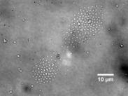
Here the rapid electrokinetic patterning technique is used to arrange bacteria into a specific pattern. The technique may be used as a tool for nanomanufacturing because it shows promise for the assembly of suspended particles, called colloids. The ability to construct objects with colloids makes it possible to create structures with particular mechanical and thermal characteristics to manufacture electronic devices and tiny mechanical parts.


Particles of different sizes can be isolated by changing the electrical frequency, and the vortex moves wherever the laser is pointed, representing a method for positioning specific types of particles for detection and analysis.

No comment yet.
Scooped by Dr. Stefan Gruenwald
Scoop.it!

3D Photografting: Laser beam used as 3D painting tool to grow biological tissue or to create micro sensors

3D Photografting: Laser beam used as 3D painting tool to grow biological tissue or to create micro sensors | Amazing Science | Scoop.it
With laser beams, molecules can be fixed at exactly the right position in a three dimensional material. The new method can be used to grow biological tissue or to create micro sensors.


"3-D-photografting" is the name of the new method. Two research teams from the Vienna University of Technology collaborated closely to develop it: Professor Jürgen Stampfl's materials science team and Professor Robert Liska's research group for macromolecular chemistry. Both research groups have already attracted considerable attention in the past, developing new kinds of 3-D-printers.


When biological tissue is grown, this method can allow the positioning of chemical signals, telling living cells where to attach. The new technique also holds promise for sensor technology: A tiny three dimensional "lab on a chip" could be created, in which accurately positioned molecules react with substances from the environment. The scientists start with a so-called hydrogel -- a material made of macromolecules, arranged in a loose meshwork. Between those molecules, large pores remain, through which other molecules or even cells can migrate. Specially selected molecules are introduced into the hydrogel meshwork, then certain points are irradiated with a laser beam. At the positions where the focused laser beam is most intense, a photochemically labile bond is broken. That way, highly reactive intermediates are created which locally attach to the hydrogel very quickly. The precision depends on the laser's lens system, at the Vienna University of Technology a resolution of 4 µm could be obtained.


Depending on the application, different molecules can be used. 3-D photografting is not only useful for bio-engineering but also for other fields, such as photovoltaics or sensor technology. In a very small space, molecules can be positioned which attach to specific chemical substances and allow their detection. A microscopic three-dimensional "lab on a chip" becomes possible.

No comment yet.